
Radiology has become an essential pillar of modern medicine, offering critical insights into human health through advanced imaging technologies. By enabling non-invasive visualization of the internal structures of the body, radiology aids in the diagnosis and treatment of various medical conditions. From the initial discovery of X-rays to the integration of artificial intelligence in imaging, the field has evolved tremendously, shaping the landscape of patient care and treatment outcomes. This comprehensive overview will explore the history, techniques, and future innovations in radiology, emphasizing its pivotal role in contemporary healthcare.
A Brief History of Radiology
The Discovery of X-rays
The journey of radiology began in 1895 with the accidental discovery of X-rays by German physicist Wilhelm Conrad Röntgen. Röntgen’s experiments with cathode rays led to the creation of an image of his wife’s hand, revealing her wedding ring and demonstrating the potential of X-rays to penetrate human tissues. This groundbreaking moment marked the beginning of diagnostic radiology and earned Röntgen the first Nobel Prize in Physics in 1901.
Early Developments
Following Röntgen’s discovery, the early 20th century saw rapid advancements in radiographic techniques. The introduction of fluoroscopy in the 1890s allowed real-time visualization of internal organs. By the 1920s, the use of radiology expanded to various medical fields, including orthopedics and oncology. However, the initial enthusiasm was tempered by concerns over radiation exposure and safety, leading to the establishment of guidelines for safe practice.
Advancements in Imaging Technology
The latter half of the 20th century brought revolutionary changes to radiology:
- Computed Tomography (CT): In the 1970s, Sir Godfrey Hounsfield and Dr. Allan Cormack developed CT scanning, which combined X-ray images taken from multiple angles to create detailed cross-sectional images of the body. This innovation greatly enhanced diagnostic accuracy, particularly in trauma and oncology.
- Magnetic Resonance Imaging (MRI): The 1980s saw the introduction of MRI, which uses strong magnetic fields and radio waves to generate high-resolution images of soft tissues without ionizing radiation. MRI has become indispensable for neurological, musculoskeletal, and cardiovascular imaging.
- Ultrasound: This imaging modality, utilizing high-frequency sound waves, gained prominence for its safety and real-time capabilities. Ultrasound is particularly valuable in obstetrics, allowing for monitoring fetal development and assessing abdominal organs.
- Nuclear Medicine: This branch employs radioactive materials to visualize metabolic processes within the body. Techniques like positron emission tomography (PET) provide insights into cancer detection and treatment response.
Techniques in Radiology
Radiology encompasses a variety of imaging techniques, each with specific applications and advantages.
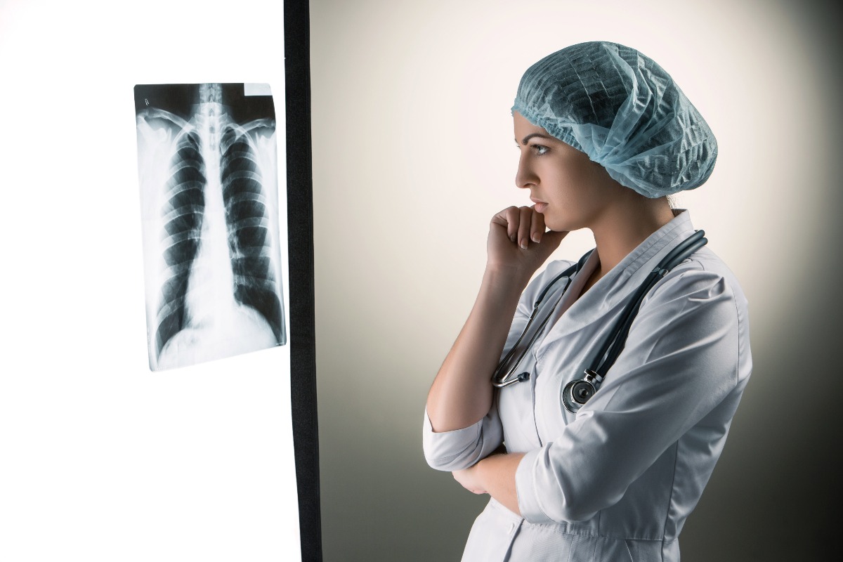
X-ray Imaging
X-ray imaging remains one of the most widely used modalities in clinical practice. It is particularly effective for diagnosing bone fractures, infections, and certain tumors. The basic principle involves passing a controlled amount of ionizing radiation through the body. Dense structures, like bones, absorb more radiation, appearing white on the resulting images, while softer tissues appear in varying shades of gray.
Applications of X-ray Imaging
- Trauma Assessment: X-rays are often the first imaging modality used in emergency settings to evaluate fractures and dislocations.
- Chest Imaging: Chest X-rays are critical for diagnosing respiratory conditions such as pneumonia, tuberculosis, and lung cancer.
- Dental Imaging: Dental X-rays provide essential information for diagnosing cavities, infections, and abnormalities in dental structures.
Computed Tomography (CT)
CT scans revolutionized the field by providing detailed cross-sectional images. This technique is invaluable for evaluating complex injuries, detecting tumors, and planning surgical interventions.
Applications of CT Imaging
- Trauma Evaluation: CT is the gold standard for assessing internal injuries in trauma patients, offering rapid and detailed views of the abdomen, pelvis, and head.
- Oncology: CT scans are crucial for tumor detection, staging, and monitoring treatment responses, helping oncologists develop effective treatment plans.
- Cardiac Imaging: CT angiography provides detailed images of blood vessels, allowing for the assessment of coronary artery disease and other vascular conditions.
Magnetic Resonance Imaging (MRI)
MRI has become the preferred imaging modality for soft tissue evaluation due to its ability to provide high-resolution images without ionizing radiation. It is particularly useful for imaging the brain, spinal cord, joints, and soft tissues.
Applications of MRI
- Neurological Disorders: MRI is essential for diagnosing conditions such as strokes, multiple sclerosis, and brain tumors, offering detailed views of brain structures.
- Musculoskeletal Imaging: MRI provides excellent visualization of ligaments, tendons, and cartilage, making it invaluable in orthopedics.
- Abdominal Imaging: MRI is increasingly used to evaluate liver, kidney, and pancreatic conditions, especially in patients who require imaging without radiation exposure.
Ultrasound
Ultrasound is a versatile imaging technique that is widely used due to its safety and real-time capabilities. It involves the emission of high-frequency sound waves, which create images based on the echoes returned from internal structures.
Applications of Ultrasound
- Obstetrics: Ultrasound is the standard method for monitoring fetal development, assessing gestational age, and detecting congenital anomalies.
- Cardiology: Echocardiography is a key tool for evaluating heart function, assessing valvular disease, and diagnosing cardiomyopathies.
- Guided Procedures: Ultrasound is frequently used to guide biopsies and aspirations, providing real-time visualization of the target area.
Nuclear Medicine
Nuclear medicine involves the administration of radioactive isotopes to visualize metabolic processes within the body. It provides functional information about organs and tissues, complementing anatomical imaging.
Applications of Nuclear Medicine
- Cancer Diagnosis: PET scans are widely used in oncology to detect tumors, evaluate metastasis, and assess treatment responses.
- Cardiac Imaging: Nuclear stress tests help evaluate blood flow to the heart muscle, assisting in the diagnosis of coronary artery disease.
- Bone Scans: Nuclear medicine is effective in detecting bone metastases and assessing conditions like osteomyelitis.
Challenges in Radiology
Despite its many advantages, radiology faces several challenges that impact its practice:
Radiation Exposure
One of the primary concerns in radiology is radiation exposure from imaging procedures. While the benefits of diagnostic imaging often outweigh the risks, minimizing radiation exposure is a priority. Radiologists adhere to the ALARA principle (As Low As Reasonably Achievable) to ensure patient safety.
Technological Advancements
The rapid pace of technological advancements in radiology necessitates ongoing education and training for radiologists and technologists. Staying current with the latest techniques, software, and imaging modalities is essential for maintaining high standards of care.
Interpretation Errors
Radiology relies heavily on the accurate interpretation of images. Misinterpretations can lead to misdiagnoses, which can have serious consequences for patient care. Continuous education, peer review, and quality assurance measures are essential to mitigate these risks.
Access to Imaging Services
Access to advanced imaging services can be limited in rural and underserved areas, creating disparities in healthcare. Efforts to expand access to imaging technology and trained specialists are crucial for improving patient outcomes.
Future Innovations in Radiology
The future of radiology is promising, with several key trends and innovations on the horizon.
Artificial Intelligence (AI)
AI is poised to transform the field of radiology by enhancing image interpretation, automating workflows, and improving diagnostic accuracy. Machine learning algorithms can analyze vast amounts of data, identifying patterns and anomalies that may be overlooked by human eyes.
- Enhanced Image Analysis: AI-powered tools can assist radiologists in detecting conditions such as tumors, fractures, and infections more accurately and efficiently.
- Workflow Optimization: AI can streamline administrative tasks, allowing radiologists to focus more on patient care and complex cases.
Teleradiology
The rise of telemedicine has facilitated the growth of teleradiology, enabling radiologists to interpret images remotely. This approach improves access to imaging services, particularly in rural and underserved areas.
- Consultation and Collaboration: Teleradiology allows for collaboration between specialists, enabling timely consultations and second opinions regardless of geographic location.
- Rapid Reporting: Remote access to imaging studies can lead to faster diagnoses and improved patient care, especially in emergency situations.
Personalized Medicine
Advancements in genomics and molecular imaging are paving the way for personalized medicine in radiology. Tailoring imaging protocols and treatment plans based on an individual’s genetic makeup and disease characteristics is becoming increasingly feasible.
- Targeted Imaging Techniques: Personalized imaging strategies can enhance the accuracy of diagnoses and treatment responses, allowing for more effective interventions.
Enhanced Imaging Techniques
Research continues to drive innovations in imaging technologies, improving resolution and functionality. Techniques such as high-resolution MRI, functional MRI (fMRI), and novel PET imaging agents are on the rise.
- Hybrid Imaging: Combining different imaging modalities, such as PET/CT or MRI/PET, allows for comprehensive evaluations of both anatomical and functional aspects of diseases.
- Real-time Imaging: Future advancements may enable real-time imaging capabilities, enhancing the ability to monitor dynamic physiological processes during procedures.
Conclusion
Radiology has become an integral component of modern medicine, providing essential tools for diagnosis, treatment, and patient care. Its rich history, diverse techniques, and continuous advancements reflect the field’s importance in improving health outcomes. As technology evolves, radiology will continue to adapt, integrating innovations like artificial intelligence and personalized medicine to enhance diagnostic accuracy and efficiency. The future of radiology is bright, promising to further shape the landscape of healthcare and patient management for years to come.